Measurement Procedures
Structural imaging


The tissue structures in the brain vary widely and posess different magnetic properties. That is, they emit signals of differing strengths when the applied electromagnetic waves induce nuclear spin. The scanner can record these signals and convert them into anatomical images of the brain. This is called structural imaging.
Repeated acquisition of structural image data over longer periods of time permits researchers to identify even small local alterations of brain anatomy. This is often done in intervention studies that examine the effects of memory or fitness training on the brain. Such studies require that MRI measurements are carried out several times, for example, before, during, and after a long-term training program. The researchers can then use the images to examine whether structural alterations of the brain are associated with the training, and to what degree. They can also investigate whether such anatomic changes are related to potential changesof cognitive performance.
Functional Imaging
Beyond structural imaging, it is also possible to watch the brain “at work.” When a certain brain region is active, it uses up oxygen. Blood coming from the lungs transports new oxygen to the active region. As oxygen-rich blood has other magnetic properties than oxygen-depleted blood, differences in the respective brain regions emerge depending on whether they are active or not. This means that brain activity during cognitive tasks can also be detected in the MR scanner. This is called functional brain imaging. In most cases, tasks are presented to participants in the scanner by visually via projection or auditorily via headphones. Working on them either activates or deactivates the functionally relevant brain regions.
Diffusion-Weighted Imaging
 and red for top-bottom connections (= superior-inferior).")
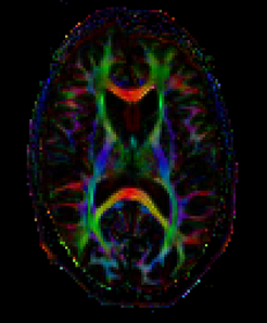
A further MRI method is used in some MPIB studies: Diffusion-weighted imaging (DWI). This makes it possible to determine the extent of diffusion movement of the water molecules in the brain’s white matter. Researchers can use this information to examine microstructural changes in the brain across the lifespan or due to targeted training, for instance.
DWI assesses the microscopic movement of water in brain tissue. As the molecules move in different directions, it is possible to measure directional profiles of this diffusion for every single image point. These profiles allow conclusions about the microstructural characteristics of the brain—weaker diffusion is explained by higher membrane density, and the main direction of the diffusion profiles in white matter is interpreted as reflecting the spatial orientation of neuronal fiber tracts.
High-Resolution Imaging of the Hippocampus


A special form of structural imaging can identify and demarcate subfields of the hippocampus using particularly high resolution and optimized contrast. The two hippocampi are located at the inner edges of the right and left frontal lobes of the brain and play a central role in processes of learning and memory. The hippocampal subfields take on specific tasks in that context.
When examining structural changes of the hippocampus within a training study, for example, determining volume changes of the hippocampal subfields in detail is of particular interest. The measurement procedure required for this purpose is optimized to demarcate the hippocampal subfields.
Magnetic Resonance Spectroscopy
")
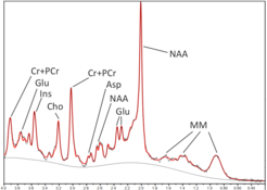
Magnetic resonance spectroscopy does not lead to the creation of images. Instead, biochemical substances in the body are assessed, allowing their concentrations to be measured within defined areas of the brain. This provides researchers with important information about the metabolism of a certain brain region, for example. The measurement of concentrations of neurotransmitters such as glutamate and glutamine in the brain is of particular interest to advance the understanding of brain processes.